Graphene oxide is a substance known for its flexibility. It’s true in a literal sense, as when it is used in solar cells or electronics, and true in the sense that it is extremely versatile.
In fact, graphene oxide — i.e., an oxidized form of graphene — can be found in such divergent items as lithium batteries, sensors and membranes. And in March 2020, an international team of scientists discovered that when that substance was 3D-printed in combination with a certain protein, it could form what were described as “tissue-like vascular structures.”
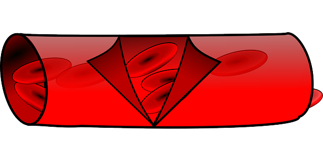
This represents the latest advance in the development of artificial blood vessels, which in time could be used to treat everything from cardiovascular disease to gunshot wounds.
The study, headed by Professor Alvaro Mata at the University of Nottingham/Queen Mary University London, concluded that the graphene oxide and protein went through a process of self-assembly, defined as the process through which various components interact to form new, functional structures. More specifically, the graphene oxide provides the framework for what Mata described in a news released as “micro-scale capillary-like fluidic structures that are compatible with cells, exhibit physiologically relevant properties, and have the capacity to withstand flow.”
Dr. Yuanhao Wu, the project’s lead researcher, noted in that same release that self-assembly on a molecular level had until now been “limited” despite the scientific community’s best efforts. This breakthrough, she added, represents a technique that “can be easily integrated with additive manufacturing to easily fabricate biofluidic devices that allow us (to) replicate key parts of human tissues and organs in the lab.”
That could have particular implications for patients undergoing bypass surgery as a result of cardiovascular disease, which according to the World Health Organization is the number one cause of death around the globe.
Research in the area of artificial blood vessels has, as a result, taken on added weight, and dates back to 1986, when scientists attempted to make them from bovine aortic cells. Particularly promising were two discoveries in 2017 — one in China, and one in the United States, at the University of California San Diego — through the use of 3D printers.
The Chinese researchers combined stem cells and nutrients to construct a graft that was used to replace a portion of an artery in the abdomens of 30 rhesus monkeys, while the U.S. scientists created a graft — subsequently used in mice — out of vascular cells that were converted into hydrogels.
Then, in 2019, researchers from Yale University and a Durham, N.C.-based medical technology firm called Humacyte used arterial cells from cadavers to create a collagen/protein structure known as a cellular matrix — i.e., the scaffolding that gives blood vessels their shape. They were then implanted in the arms of patients suffering from kidney disease, and over time came to be populated by the patients’ own cells.
The discovery in March of this year represents the next step in the creation of artificial blood vessels, and may someday prove to be a giant leap forward. Given the prevalence of cardiovascular disease, it is impossible to overstate the importance of such work, and what it might mean in the future.

